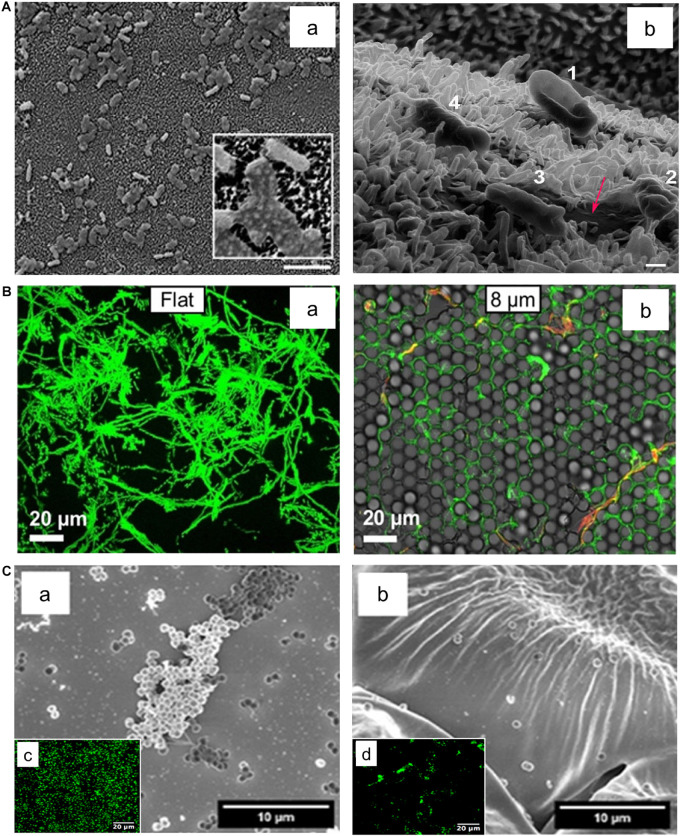
FIGURE 3.
Effect of surface topography on bacterial adhesion. (A) E. coli bacteria attached on textured surface of dragonfly wing (a). The Helium ion microscopy image (b) indicate progressive dying stages of E. coli on the dragonfly wing starting with No. 1 where the cell attached to the surface and its membrane deforms, ending with No. 4 where the cell membrane lost its integrity and cell sank into nanopillars. Scale bar is 200 nm. Adapted with permission from Bandara et al. (2017) Bactericidal Effects of Natural Nanotopography of Dragonfly Wing on Escherichia coli. ACS Appl Mater Interfaces 9, 6,746–6,760. Copyright © 2017, American Chemical Society. (B) Comparison of P. aeruginosa adhesion on flat (a) and textured surfaces (b). P. aeruginosa appears to cover a smaller surface area on the textured surface with features in 8 μm range in comparison to the flat surface. Adapted with permission from Chang et al. (2018) Surface Topography Hinders Bacterial Surface Motility. ACS Appl Mater Interfaces 10, 9,225–9,234. Copyright © 2018, American Chemical Society. (C) Adherence of S. epidermidis on flat (a) and rose petal-textured (b) surfaces after 2 h incubation. SEM (a,b) and fluorescence microscopy (c,d) images of S. epidermidis showed a decreased amount of bacterial adhesion on the textured surface. Adapted with permission from Cao et al. (2019) Hierarchical Rose Petal Surfaces Delay the Early-Stage Bacterial Biofilm Growth. Langmuir 35, 14,670–14,680. Copyright © 2019, American Chemical Society.

