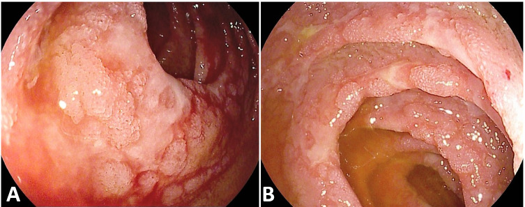
Figure 3.
Small intestine. A patient without medical history or chronic therapy, admitted in a non-intensive ward, underwent a CT scan for persistent abdominal pain, showing thickened jejunal walls; enteroscopy performed 44 days after admission showed multiple erosions and ulcers on a background atrophic mucosa with shortened villi. Histology reported acute inflammation, ulcerations and granulation tissue, with abundant eosinophils, without definite aetiology (all cultural exams negative including immunohistochemistry for Cytomegalovirus and PCR for SARS-CoV-2).

