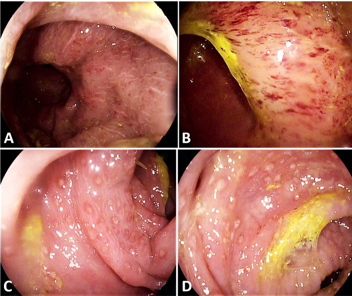
Figure 7.
Colonoscopy of a young patient performed for persisting diarrhoea 2 months after being discharged from a COVID-19 ward. In the rectum, a fragile and dystrophic mucosa with diffuse petechiae was seen (A, B). The mucosa of the distal sigmoid colon appeared severely oedematous, congested, with diffuse aphthous erosions (C, D). Histology of the whole colon showed intense lymphocytic and granulomatous infiltration.

