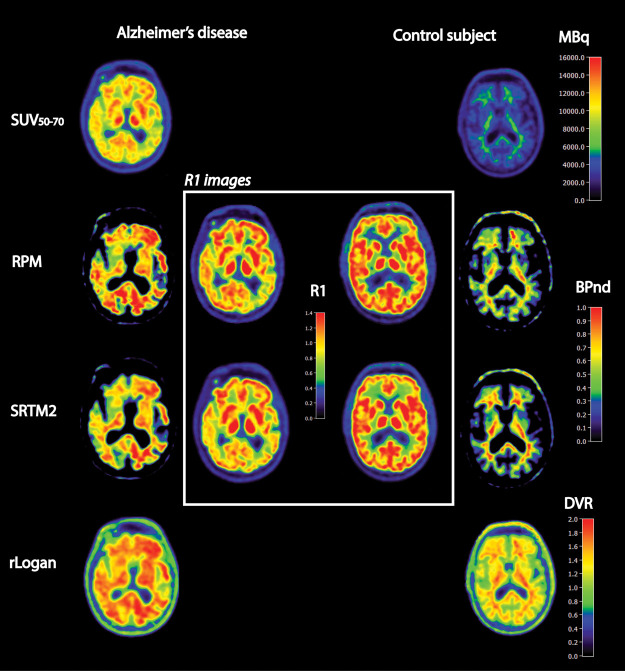
Figure 1.
Examples of several quantitative images of a selection of parametric methods for a typical Alzheimer’s disease subject and a healthy volunteer. If available (RPM, SRTM2), we also presented (in the center white box) the corresponding R1 images reflecting tracer delivery or relative cerebral blood flow.

