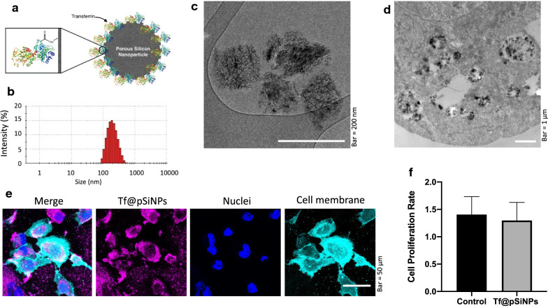
Fig. 1.
Tf@pSiNPs characterisation and uptake. a Representation of Tf@pSiNPs (not to scale). Transferrin structure obtained from Protein Data Bank [38]. b Hydrodynamic particle size distribution of Tf@pSiNPs, as indicate by DLS. c Cryo-TEM image of Tf@pSiNPs d Cryo-TEM image of Tf@pSiNPs in glioma cells. e Confocal microscopy imaging to show Tf@pSiNPs’ uptake by WK1 cells. Cyanine5 (magenta), Vybrant (cyan) and Hoechst 33,342 (blue) staining allowed visualisation of Tf@pSiNPs, cell membrane and nuclei, respectively. f WK1 cell proliferation rate following treatment with Tf@pSiNPs, quantified as the ratio between the number of cells 24 and 48 h post-seeding (data presented as mean ± 1 SD, n = 3), no significant difference was found following treatment with Tf@pSiNPs (Student’s t-test). Tf@pSiNPs transferrin-functionalised porous silicon nanoparticles, DLS dynamic light scattering, TEM transmission electron microscope

