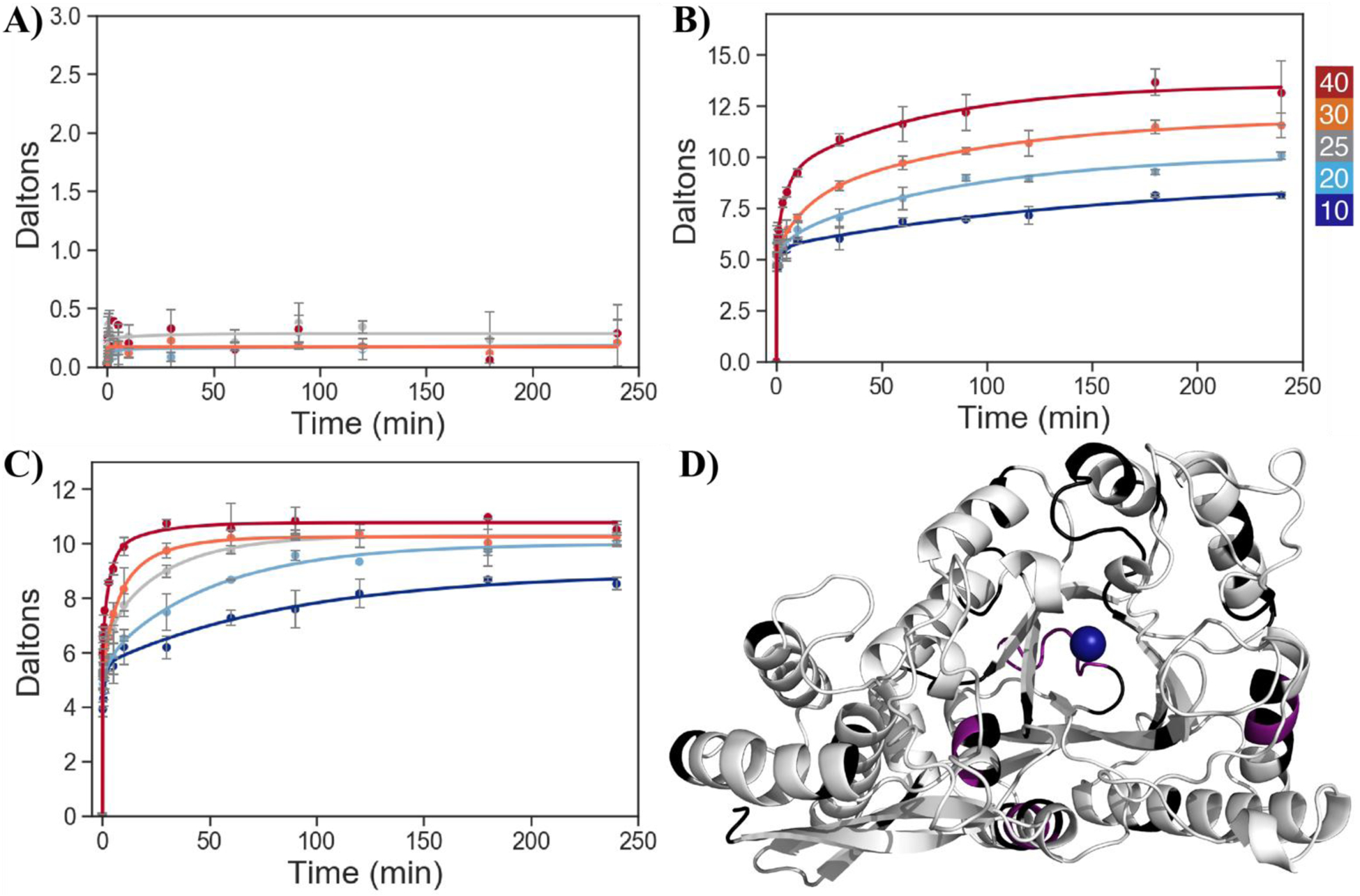
Figure 6.

Representative HDX uptake plots showing the time dependence of peptide-specific deuteration at different temperatures for WT peptides in the open structure; color scale for temperature at top right. A) 224–228, Type I, which shows temperature-independent exchange in the HDX experimental timescale, B) 263–279, Type II, where the different temperatures approach a common plateau, and C) 94–112, Type II, where HDX traces do not converge even at the longest times and high temperature. D) Crystal structure of holo-enolase with Type I, Type II, and non-reporting regions shown in violet, white, and black, respectively. The tightly bound magnesium ion is shown as a blue sphere. Yeast enolase is a homodimer with a C2 axis of symmetry; for the orientation of the monomer above, the dimer interface is horizontally located along the very bottom of the subunit. PDB 1EBH.
