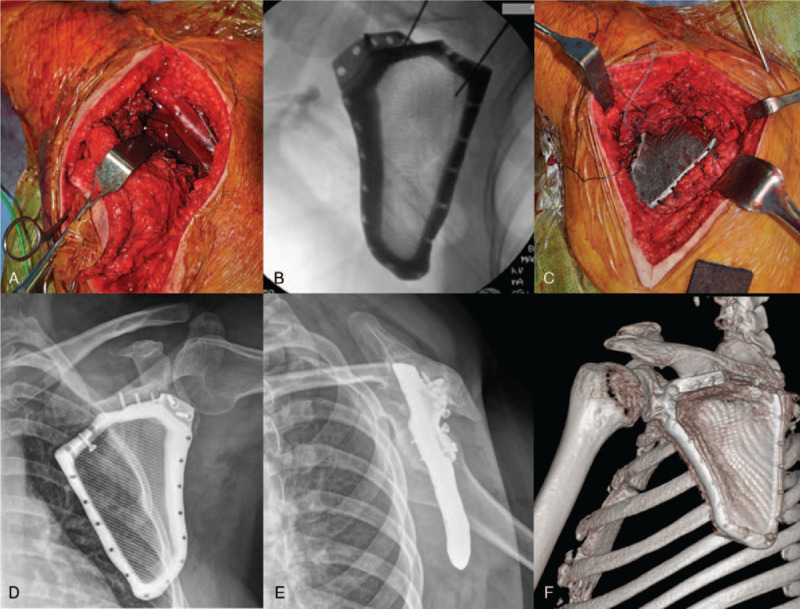
Figure 3.

The osteotomy guide to the spine of the scapula and removal of the infrascapular area (A). After implant fixation to the retained scapula by using intraoperative fluoroscopy (B), the surrounding muscle tissue is sutured to the prosthesis through the holes at the edge of the prosthesis (C). The postoperative plain radiographs (D and E) and computed tomography images (F) show good match between the printed segmental scapula prosthesis and the retained scapula.
