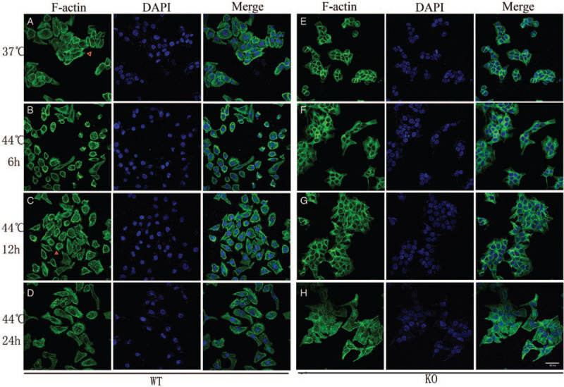
Figure 1.

Immunofluorescence staining of F-actin (original magnification ×400). Cells were heated at 44.0°C for 30 min and allowed to recover under normal growing condition for up to 6, 12, and 24 h (original magnification ×400). At different time intervals, the cells were stained and processed for scanning confocal microscopy. (A) Unheated WT cells; (B–D) Hyperthermia treatment of WT cells, (B) 6 h, (C) 12 h, (D) 24 h after hyperthermia. (E) Unheated DnaJA4-KO cells; (F–H) hyperthermia treatment of DnaJA4-KO cells, (F) 6 h, (G)12 h, (H) 24 h after hyperthermia. F-actin (phalloidin) shown in green and nuclei (DAPI) shown in blue. WT: Wild-type; DnaJA4-KO: DnaJA4-knockout; DAPI: 4′,6-diamidino-2-phenylindole.
