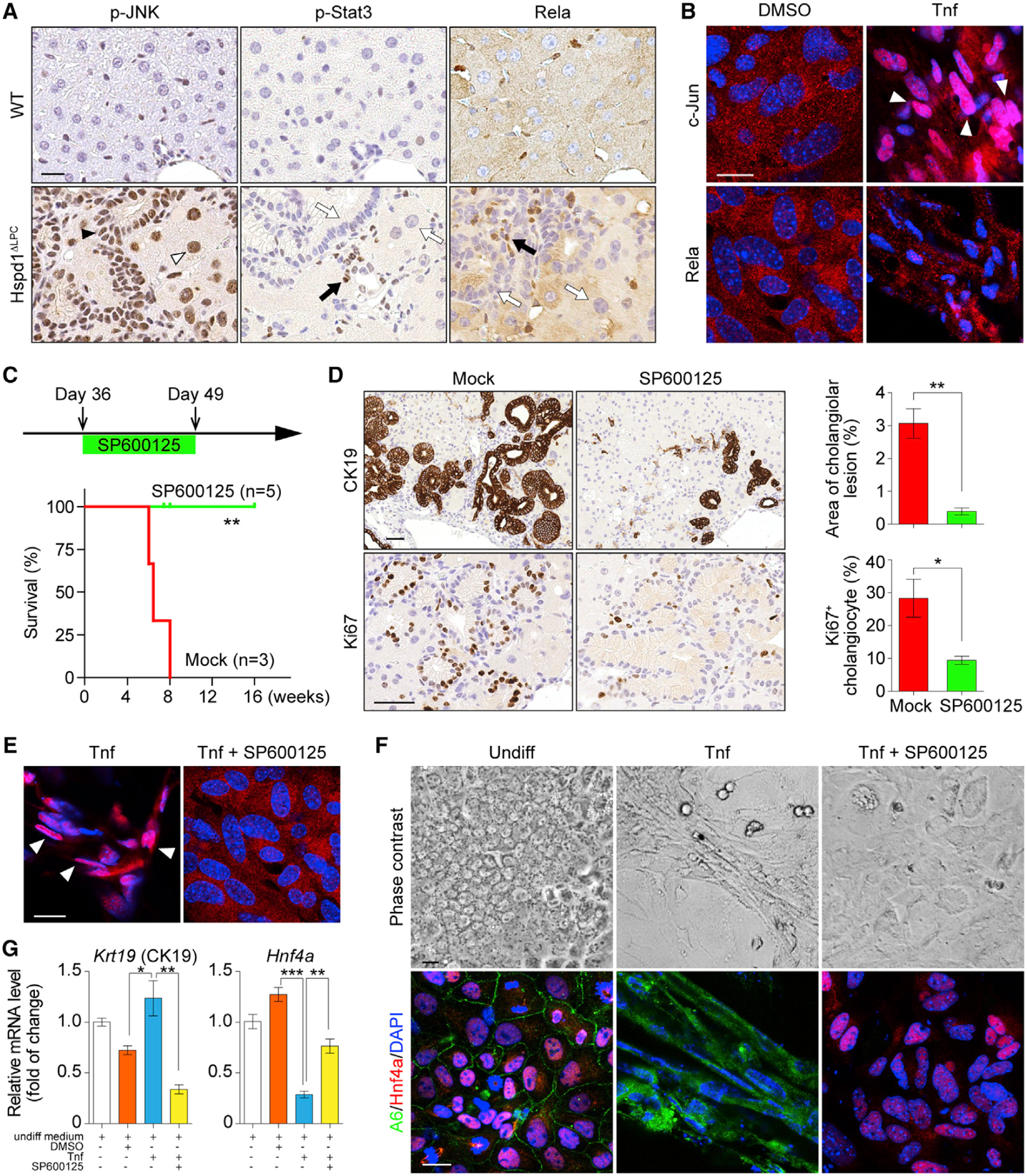
Figure 6. JNK/c-Jun Activation Is Required for Premalignant Cholangiocellular Lesions.

(A) p-JNK, p-Stat3, and Rela IHC in 8-week-old WT and Hspd1ΔLPC livers. The black arrowhead indicates a p-JNK+ cholangiocyte, the white arrowhead indicates a p-JNK+ hepatocyte. Black arrows indicate positive staining in non-parenchymal cells, while white arrows indicate negative staining in liver parenchymal cells. Scale bar, 20 μm.
(B) Rela and c-Jun IF in hepatoblasts cultured in basal medium supplemented with DMSO or Tnf. White arrowheads indicate c-Jun nuclear staining. Scale bar, 20 μm.
(C) Timeline of SP600125 administration on Hspd1ΔLPC mice and survival of Hspd1ΔLPC mice not treated (mock) or treated with SP600125.
(D) CK19 and Ki67 IHC in 8-week-old Hspd1ΔLPC livers treated with mock or SP600125, and quantification of cholangiolar cancerous lesion areas and Ki67+ cholangiocytes relative to total cholangiocytes. Scale bar, 50 μm.
(E) c-Jun IF in hepatoblasts cultured in Tnf-containing medium with or without SP600125. White arrowheads indicate the c-Jun nuclear staining. Scale bar, 20 μm.
(F and G) Phase contrast and A6 and Hnf4α IF images (F) and qRT-PCR (G) of hepatoblasts in basal or Tnf-containing medium with or without SP600125. Scale bar, 20 μm.
Data are represented as the mean ± SEM. *p < 0.05, **p < 0.01, ***p < 0.001. ns, not significant. See also Figure S6.
