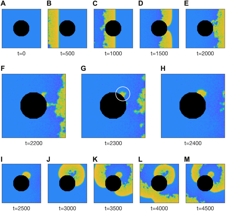
Fig 8. Ectopic activity induced at the tissue boundary by a passing wave.
The 100 × 100 medium is briefly stimulated at the left-hand boundary and the ensuing wave propagates to the right. The black region represents the tissue boundary. The white circle in panel G marks the genesis of ectopic activity by cells on the tissue boundary that fail to repolarize. The medium is densely heterogeneous with parameter b of each cell drawn randomly from a log-normal distribution (μ = 1, σ = 1). The coupling coefficient is c = 0.7. See S3 Video for an animated version of this figure.

