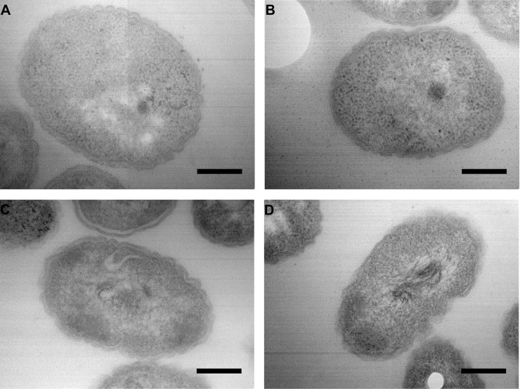
Fig 3. Microscopic observations by TEM (transmission electron microscopy).
Observation of the three strains isolated in 2014 and 2016 (A: strain A2; B: strain B1; C: strain C), and reference strain (D: strain K96243). The scale bar represents 250 nm. The observations presented here are without trimethoprim induction, the same pictures were obtained with trimethoprim induction.

