Fig. 1.

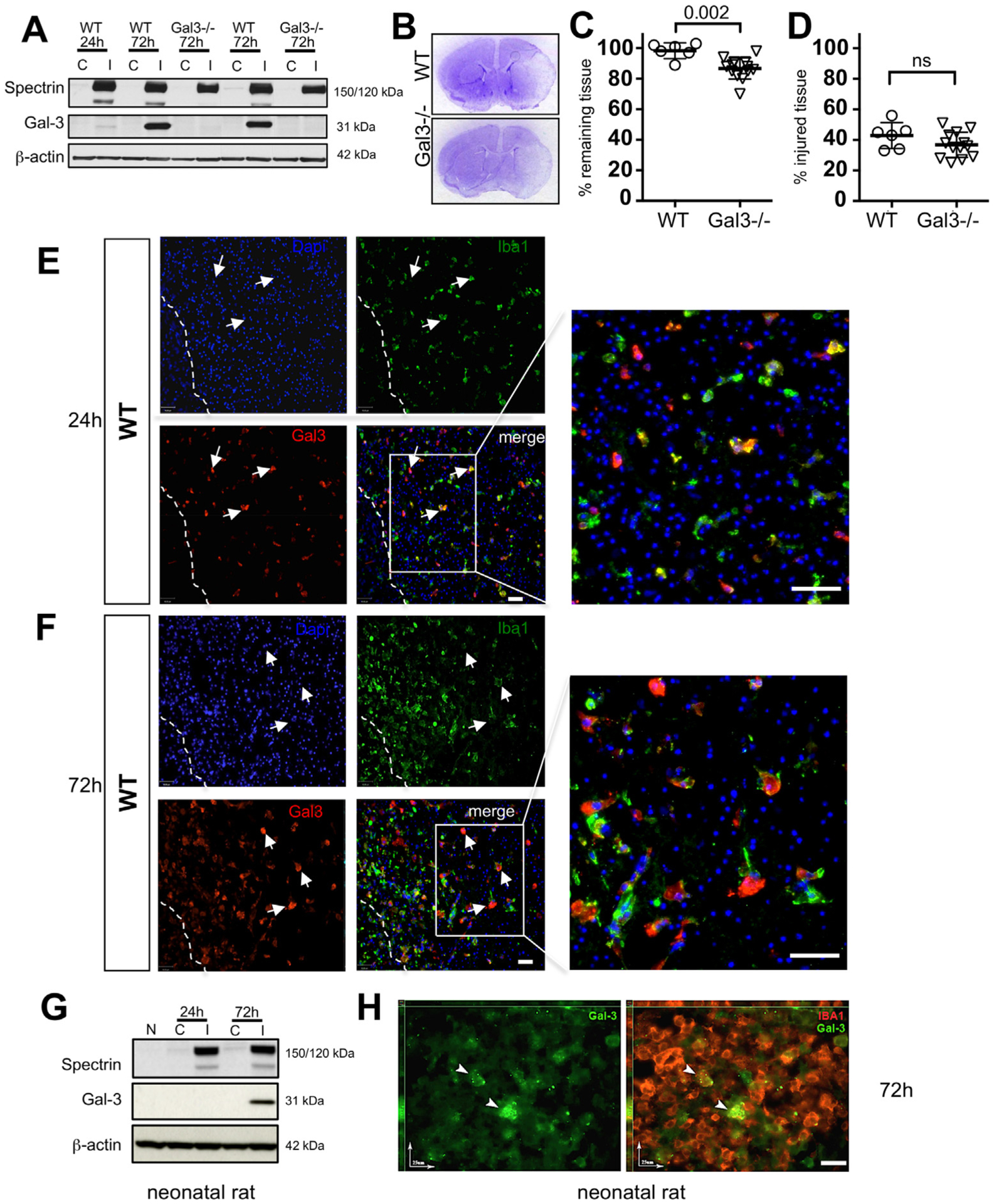
Gal3 is upregulated after tMCAO in neonatal mice and worsens injury. A. Top row: A representative Western Blot that demonstrates the presence of spectrin cleavage by calpain (150 kDa) and caspase-3 (120 kDa)-dependent mechanisms in injured (I) regions as compared to lack of cleavage in matching contralateral (C) region. Middle row: gradual upregulation of Gal3 expression in injured region of WT and not in Gal3−/− 72 h post-MCAO. Data are normalized to beta-actin protein expression (bottom row). B. Representative Nissl stained coronal sections of WT and Gal3−/− 72 h after MCAO C. Percent volume of remaining tissue in the ipsilateral hemisphere compared to that in contralateral hemisphere. D. Percent volume of injured tissue within the ipsilateral hemisphere. E–F. In WT mice, Gal3 is expressed in injured caudate 24 h and 72 h post-tMCAO. Injured and dying cells are identified by punctate nuclei stained with DAPI. Dashed line represents peri-infarct region at 72 h post-MCAO. Arrows indicate Gal3+ activated microglia and macrophages (scale bar: 40 μm). G. A representative Western Blot of spectrin cleavage (top), Gal3 expression (middle), and β-actin (bottom) expression in the rat brain. Shown is expression in naïve (N), contralateral (C) and injured (I) regions. H. Gal3 is upregulated in a subpopulation of Iba1+ microglial cells in injured rat brain, as evident from immunofluorescence at 72 h post-tMCAO (scale bar: 25 μm). Dots in C–D represent data from individual animals (n = 6 for WT, n = 14 for Gal3−/−). Mean ± SD. Significance levels as indicated on individual panels, ns: non-significant (Student’s t-test).
