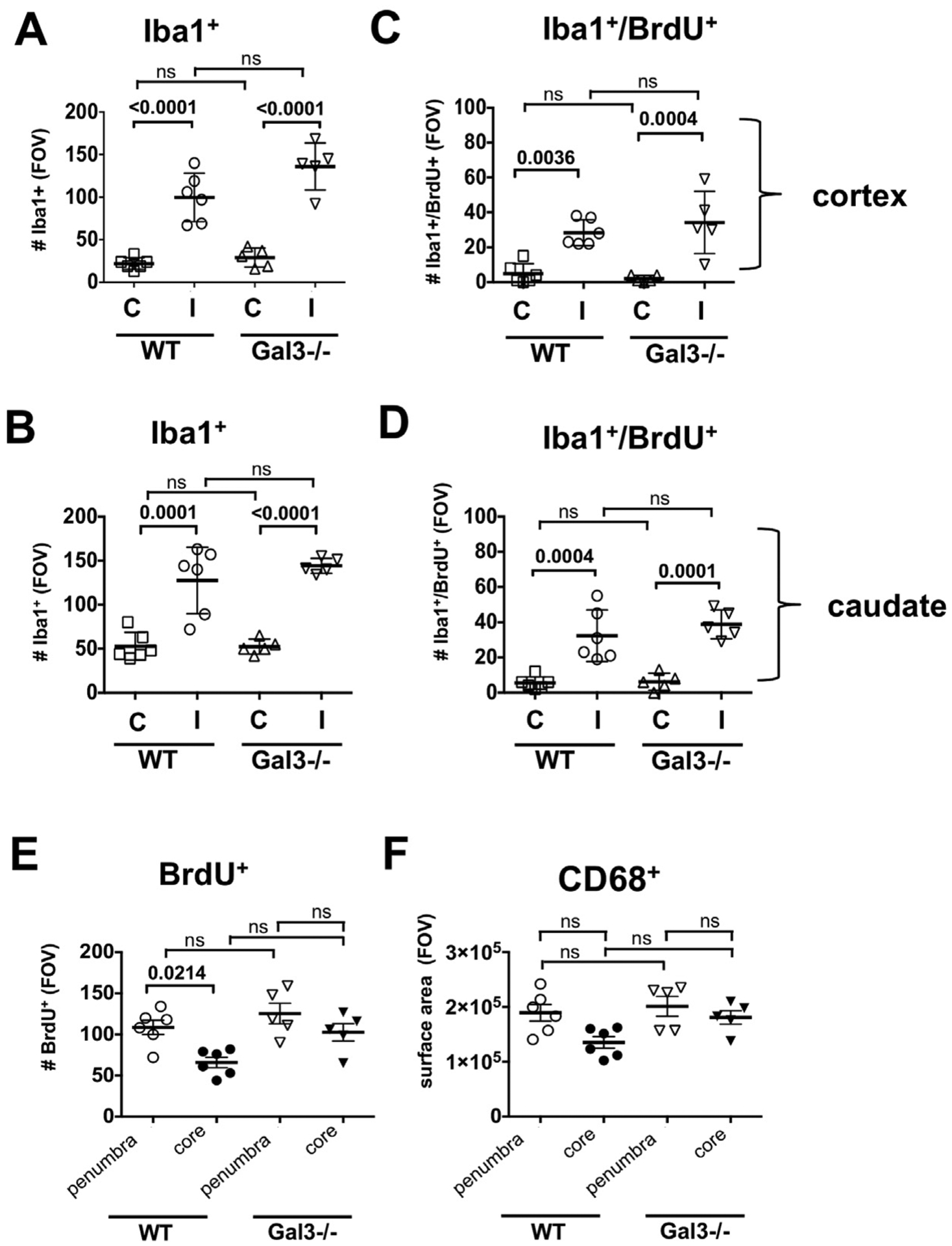
Fig. 3.

Microglial accumulation and proliferation are similar in injured cortex and caudate of Gal3−/− and WT mice 72h post-tMCAO. A–B. The number of Iba1+ in the cortex (A) and the caudate (B). C–D. Proliferation of Iba1+ cells in the cortex (C) and the caudate (D). E. The overall cell proliferation (BrdU+ cells) in the peri-focal injury and in the core. F. Acquisition of CD68+ cells in the peri-focal injury and in the core. FOV = 2.9 × 106 μm3. Dots in A–F represent data from individual animals (n = 6 for WT, n = 5 for Gal3−/−). Mean ± SD. Significance levels as indicated on individual panels, ns: non-significant (ANOVA with Bonferroni post hoc test). (C) – contralateral, (I) –injured.
