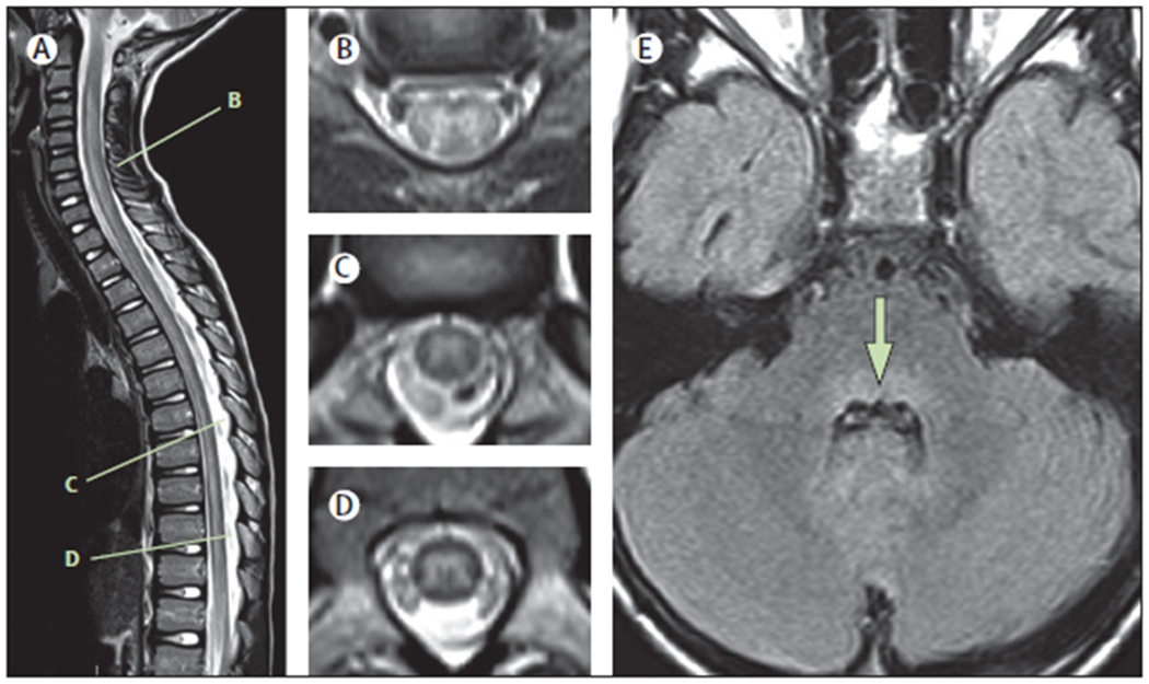
Figure 1: Typical MRI findings in the acute phase of AFM.

Spinal MRIs are shown of an 8-year-old child with AFM, acquired 24 h after onset of neurological symptoms.(A) Sagittal T2 image showing an ill-defined longitudinally extensive central/anterior spinal cord lesion. (B) Axial T2 image from C5–C6 shows hyperintensity of the entire grey matter of the spinal cord, with associated oedema and some surrounding white matter hyperintensity. (C) Axial T2 image from T7 shows asymmetric hyperintensity of the grey matter (right more than left). (D) Axial T2 image from T10 shows hyperintensity of the entire grey matter. (E) Axial FLAIR image at the level of the middle cerebellar peduncle demonstrates hyperintensity of the dorsal pons (arrow). AFM=acute flaccid myelitis.
