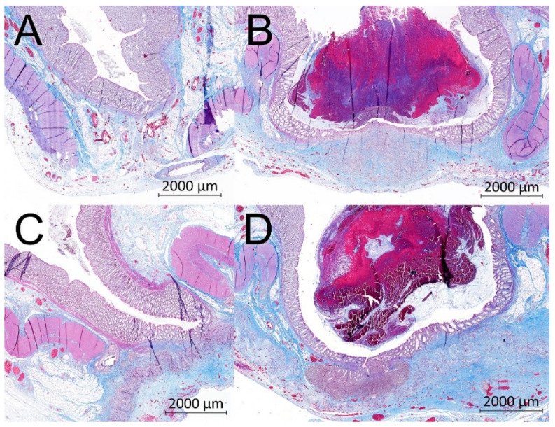
Figure 8.
Example histological specimens from both groups. Gomori trichrome (A) Control group, optimal healing, normal morphology of the intestinal wall, muscular layer with normal scar tissue; (B) Control group, larger defect of the muscular layer, a pseudodiverticulus; (C) Experimental group, optimal healing, normal morphology of the intestinal wall, visible residues of the nanofibrous material in the bottom of the image; (D) Experimental group, large defect of the muscular layer, a pseudodiverticulus, visible residues of the nanofibrous material in the bottom of the image covering the incomplete defect of the intestinal wall.

