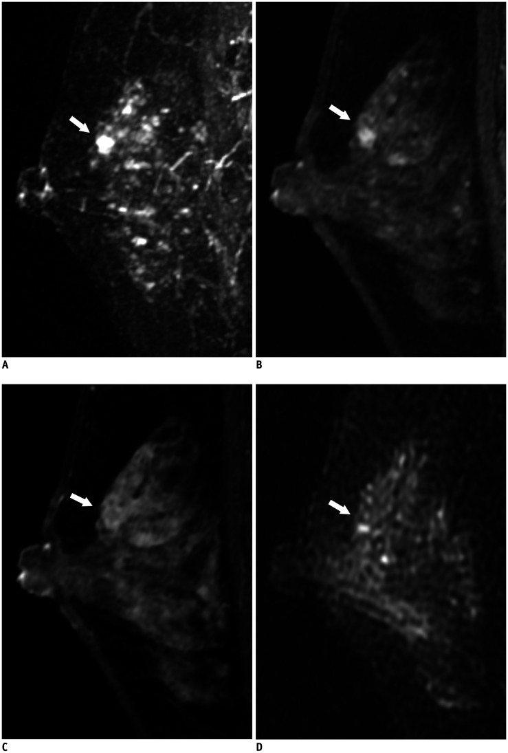
Fig. 3. True-negative lesion on abbreviated MRI in a 49-year-old woman.
MIP reconstruction image (A) and first post-contrast T1-weighted sagittal image (B) show a 0.5 cm irregular, circumscribed mass (arrows) in the left upper breast. The fifth post-contrast T1-weighted sagittal image (C) shows the mass (arrow) with clear washout kinetics. T2-weighted sagittal image (D) shows the mass (arrow) with iso-signal intensity. This lesion was classified as BI-RADS final assessment category 2 or 3 by all five radiologists on abbreviated breast MRI and category 4 by four of five radiologists on full diagnostic MRI. It was finally confirmed as sclerosing adenosis after excision.

