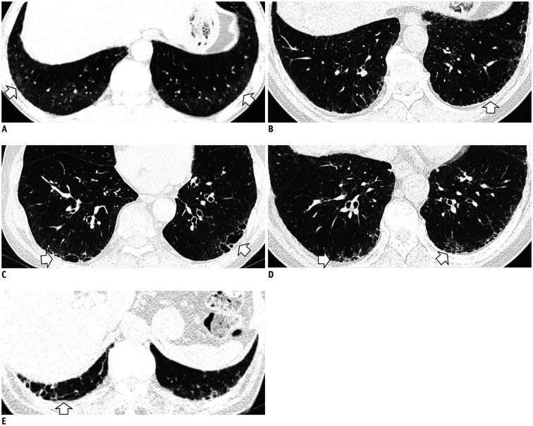
Fig. 1. Typical image patterns demonstrating ILA.
A. A representative axial CT image showing bilateral subpleural GGA (arrows). To confirm a dependent opacity, this CT was taken in a prone position. B. CT image taken in the prone position shows bilateral subpleural reticulation in both lungs (arrow). C. Subpleural non-emphysematous cysts in both lungs. These present as thin-walled variably sized cysts on CT images (arrows). D. CT images demonstrate honeycombing in both lungs (arrows). These are clustered cystic air spaces, and their sizes are usually uniform (3–10 mm). E. Image shows traction bronchiectasis/bronchiolectasis (arrow). This can be seen as thick-walled bronchial or bronchiolar dilatation in the subpleural area of the lower lobe. GGA = ground-glass attenuation, ILA = interstitial lung abnormality

