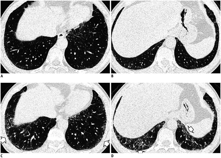
Fig. 10. Serial chest CT images show a progression of nonfibrotic ILA in a 57-year-old male former smoker with ten pack-years of cigarette consumption.
A, B. Initial CT images show mild reticular opacity in the subpleural zone. At this time, his lung function was within a normal range. C, D. Follow-up CT images after three years show the progression of reticular opacity and GGA in the peripheral portion of the lower lung (arrows). During this period, the patient experienced a decrease in total lung capacity (79% of predicted) and diffusion capacity (85% of predicted).

