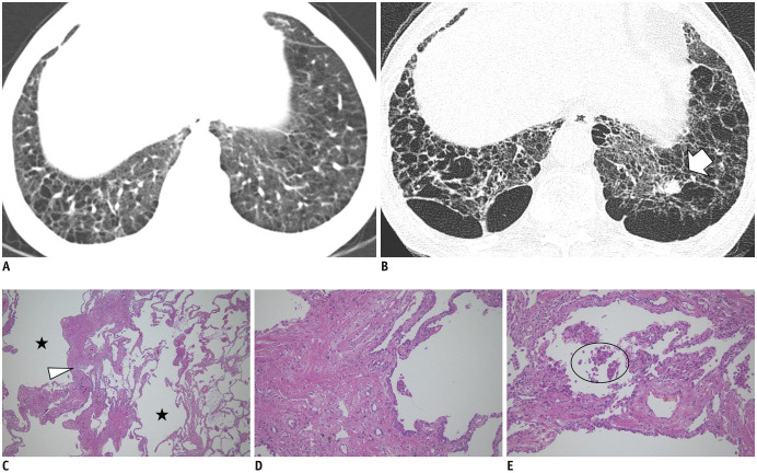
Fig. 11. Chest CT images and histopathologic findings of nonemphysematous cysts in a 60-year-old current smoker.
A. The initial image shows emphysema and nonemphysematous cysts in the lower lobes. B. After 12 years, the CT image shows the progression of emphysema and nonemphysematous cysts, with a newly developed lung nodule in the left lower lobe (arrow), which was proven to be a squamous cell carcinoma by left lower lobectomy. C. Low magnification (× 40) of the histopathologic finding shows marked interstitial thickening (arrowhead) with prominent emphysema (asterisks). D, E. High magnification (× 100) findings show interstitial thickening consists of thick collagen bundles mixed with hyperplastic smooth muscle fibers and pigmented macrophages (circle) are present in air spaces. The histopathologic findings are consistent with smoking-related interstitial fibrosis, which is a pathologic subtype of ILAs (hematoxylin & eosin staining).

