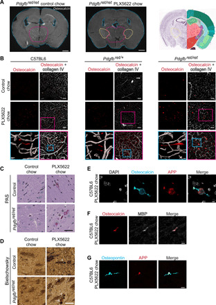
Fig. 6. Microglia ablation leads to bone protein–containing axonal spheroids in white matter.

(A) Coronal sections of Pdgfbret/ret mouse brains fed with control chow or chow containing PLX5622. Mice treated with PLX5622 exhibited conspicuous osteocalcin staining (in white) in the internal capsule (yellow dotted lines) in addition to calcifications in the thalamus (encircled with pink dotted lines). Coronal mouse brain section (Allen Adult Mouse Brain Atlas) depicts the location of analyzed brain sections (photo credit: Allen Institute). (B) Linear inclusions, positive for osteocalcin (in red), in white matter are not vessel associated in C57BL6 (left), Pdgfbret/wt (middle), and Pdgfbret/ret (right) mice. Blood vessels are visualized using the anti–collagen IV antibody (in white). (C) White matter deposits appearing in Pdgfbret/ret and control mice (Pdgfbret/wt) after PLX5622 treatment are positive for PAS (black arrowheads). (D) Dystrophic neurites exhibiting axonal spheroids (indicated by white arrowheads) in white matter after PLX5622 treatment in Pdgfbret/ret and control (Pdgfbret/wt) mice. (E to G) White matter deposits appearing after microglial depletion are positive for osteocalcin (in cyan) and APP (in red) (E), osteocalcin (in red) and MBP (in white) (F), and OPN (in cyan) and APP (in red) (G). Nuclei were visualized using DAPI (E, in white). C57BL6 control chow and PLX5622 chow, n = 5. Pdgfbret/ret and Pdgfbret/wt control chow, n = 4; and PLX5622 chow, n = 3. Scale bars, 1000 μm (A), 100 μm (B), 50 μm (B pink inset, C, and D), 10 μm (B blue inset, F, and G), and 5 μm (E).
