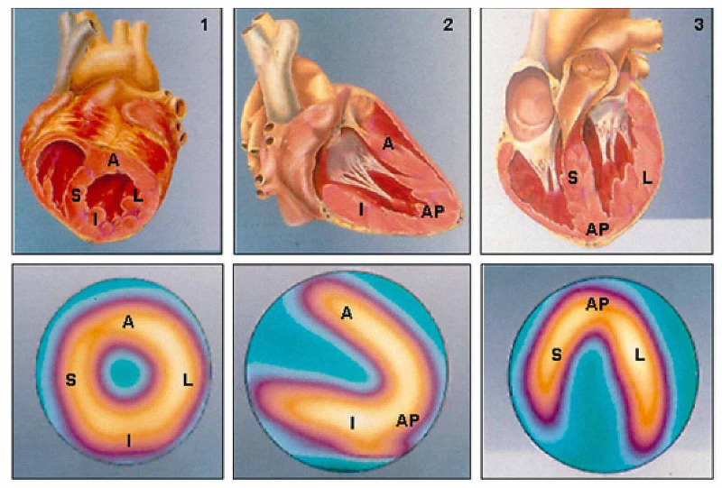
Figure 3. Two-dimensional reconstructive images of scintigraphy (lower images) and its correspondence to three-dimensional heart model (upper images).
Two-dimensional reconstruction of scintigraphy images representing normal perfusion patterns (lower images), in line with the minor axis (1), vertical long axis (2), and horizontal long axis (3) cross-sections and their respective corresponding anatomical cross-sections (upper images)
A: anterior; AP: apical; I: inferior; L: lateral; S: septal
Adapted from Mastrocola LE [24]

