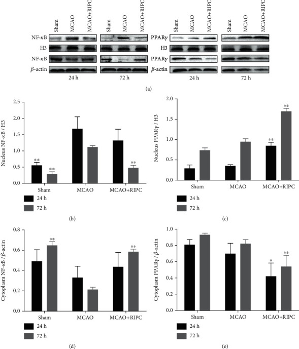
Figure 6.

RIPC repressed NF-κB shifting from the cytoplasm to nucleus and stimulated PPARγ shifting from the nucleus to cytoplasm in ischemic brain at 24 h or 72 h after MCAO. Western blot of the nucleus NF-κB (a), cytoplasm NF-κB (b), nucleus PPAR-γ (c), and cytoplasm PPAR-γ (d) in ischemic brain. Data were shown as mean ± SD (n = 3). ∗P < 0.05 and ∗∗P < 0.01 vs. the MCAO group.
