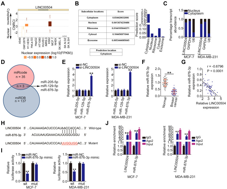
Figure 2.
LINC00504 functions as a miR-876-3p sponge in breast cancer. (A and B) lncLocator and lncATLAS were used to predict the subcellular location of LINC00504. (C) The subcellular distribution of LINC00504 in MCF-7 and MDA-MB-231 cells was determined by subcellular fractionation followed by qRT-PCR analysis. (D) Schematic diagram indicating the predicted miRNAs targeting LINC00504. (E) qRT-PCR was used to detect the expression of miR-205-5p, miR-129-5p, and miR-876-3p in MCF-7 and MDA-MB-231 cells after LINC00504 depletion. (F) MiR-876-3p expression in 57 pairs of breast cancer tissues and corresponding adjacent normal tissues as analyzed by qRT-PCR. (G) The relationship between miR-876-3p and LINC00504 expression in 57 breast cancer tissues as determined by Pearson’s correlation analysis. (H) The binding sites between LINC00504 and miR-876-3p were predicted by bioinformatics analysis. (I) Luciferase reporter assay was conducted to study the molecular interaction of LINC00504 and miR-876-3p in breast cancer cells. MCF-7 and MDA-MB-231 cells were co-transfected with LINC00504-wt or LINC00504-mut and miR-876-3p mimic or miR-NC, followed by the evaluation of luciferase activity at 48 h post-transfection. (J) RIP assay was carried out in MCF-7 and MDA-MB-231 cells, followed by qRT-PCR to measure the enrichment of LINC00504 and miR-876-3p associated with Ago2. **P < 0.01.

