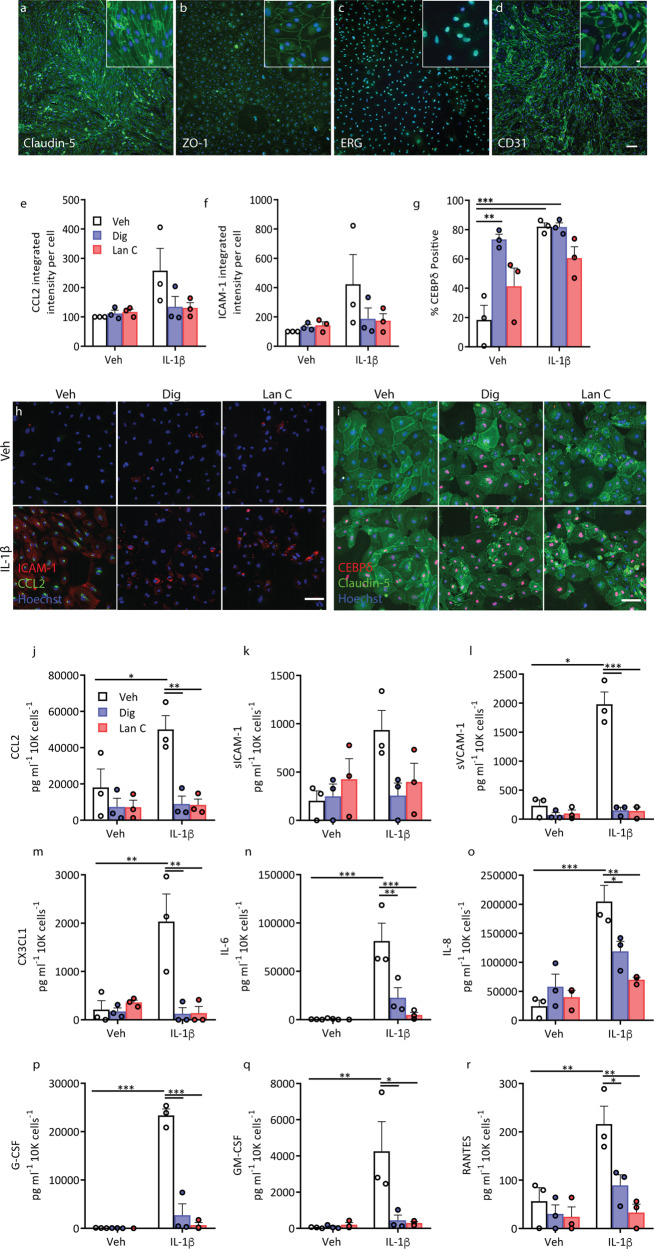
Fig. 4. Inflammatory cytokine secretion is blocked by both digoxin and lanatoside C in primary human brain endothelial cells.
Endothelial cells derived from human brain tissue express endothelial markers, Claudin-5 (a), ZO-1, (b), ERG (c) and CD31 (d). e–g Human endothelial cells were pretreated with vehicle (white bars), digoxin (100 nM, blue bars) or lanatoside C (1 µM, red bars) for 24 h, followed by 24 h of IL-1β (0.05 ng mL−1) treatment. Staining quantification of CCL2 (e), ICAM-1 (f) and nuclear CEBPδ (g), in endothelial cells (n = 3, cases used: SS42, SS50, E214). Representative images (h, i). j–r Conditioned media was analysed by CBA in endothelial cells treated as above (n = 3, cases used: SS42, SS50, E214). Cytokine/chemokine concentration was normalised to total cell counts (Hoechst)). Two-way ANOVA with Tukey’s multiple comparison test mean ± SEM. ***p < 0.001, **p < 0.01, *p < 0.05.

