Fig. 2.
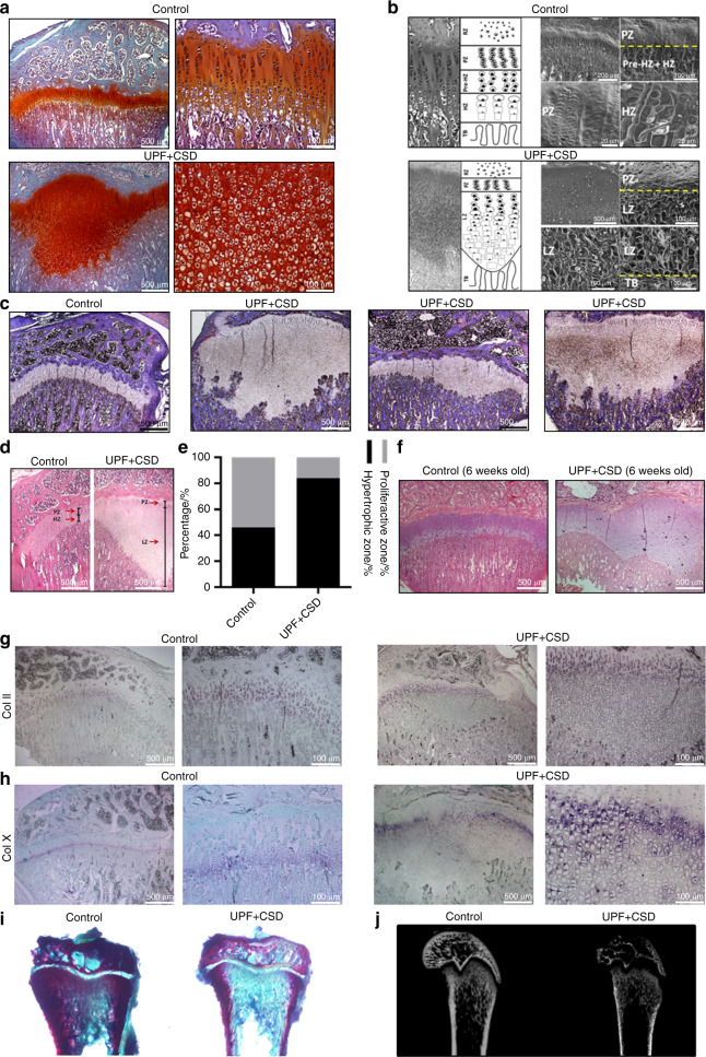
Consumption of an ultra-processed diet results in a damaged growth plate (GP) and modified chondrocyte differentiation process. Tibiae from the control and UPF + CSD groups (see legend to Fig. 1) were dissected, processed, embedded in paraffin blocks and stained with (a) safranin O, (c) Masson’s trichrome or (d and f) hematoxylin and eosin. b Light microscopy and schematic presentation (left) and surface scanning by electron microscopy (right) of the different GP zones. Resting zone (RZ), proliferative zone (PZ), prehypertrophic zone (Pre-HZ), hypertrophic zone (HZ), lesion zone (LZ), trabeculae (TB). c Masson’s trichrome staining demonstrates a variety of GP lesions. d Hematoxylin and eosin staining of the control and UPF + CSD groups. The different zones are depicted by arrows. e Quantification of the relative ratio of the zones in the GP: the widths of the whole GP and the PZ, HZ and LZ were measured at 10 different points along the GP and averaged with measurements from 6 other GP samples in each group. The percentages of PZ and HZ/LZ from the whole GP were calculated. f GP lesion at 6 weeks of age. g, h In situ hybridization analysis of Col II (top) and Col X (bottom) mRNA signals. i Alizarin red and alcian blue staining of nondecalcified tibial bone. j 2D X-ray image of femora bone

