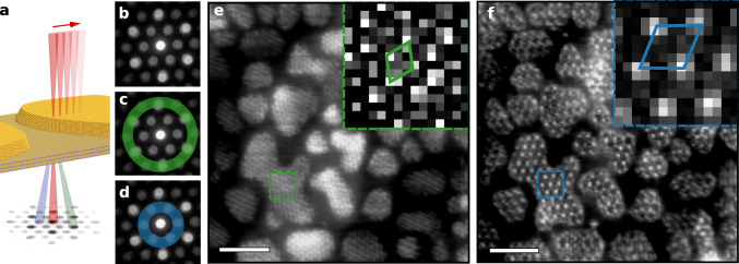
Fig. 4. 4D STEM imaging of 18 Å and 32 Å moiré periodicities.
a Schematic of 4D STEM technique showing rastered beam (red) on MoS2/Au{111} with corresponding CBED pattern at each point. The green and blue scattered beams are centred on the spots of the annuli shown in (c and d). b CBED pattern formed by averaging patterns collected over the entire scan area, c 4D STEM annulus used to isolate 18 Å moiré periodicity (angular range 31–43 mrad, green) and d 4D STEM annulus used to isolate 32 Å periodicity (angular range 11–24 mrad, blue). e, f Virtual ADF STEM images revealing 18 (green) and 32 Å (blue) period moirés, respectively. Scale bar, 200 Å. Insets show unit cells.

