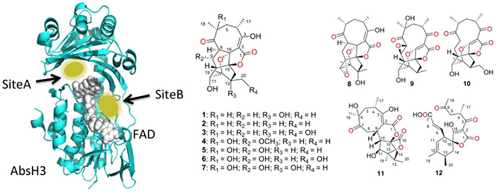
Figure 2.
Binding sites from docking studies and abyssomicin analogs used in the docking calculations. At left, FAD model showing the two sites where abyssomicin analogues bound in the docking study. Site A is the putative active site, while Site B is in the FAD binding groove. The presence of this second binding site may allow substrates to diffuse across the enzyme surface to the active site, even when not recruited directly to Site A. At right, chemical structures of the abyssomicin analogues used in the docking computations. Interestingly, compounds 8-12 preferred Site B to Site A, which is the active site.

