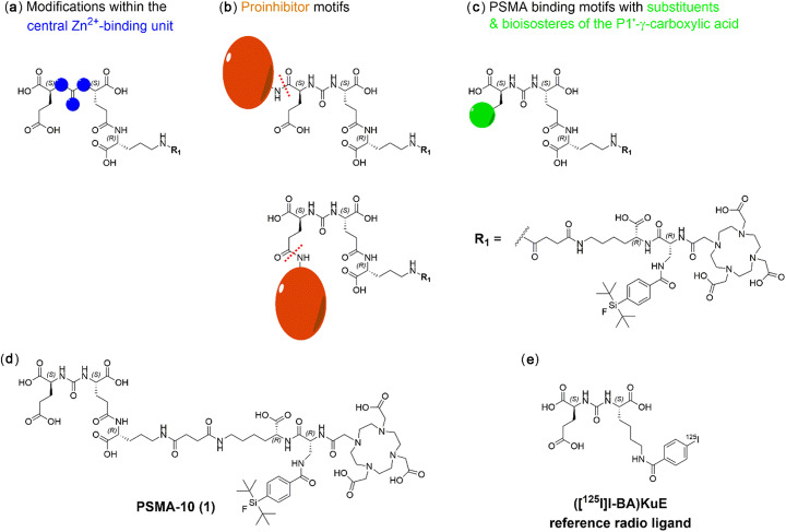
Fig. 1.
Schematic representation of PSMA inhibitors containing (a) modifications within the central Zn2+-binding unit (b) proinhibitor motifs (expected cleavage sites are indicated as red dotted lines) and (c) substituents & bioisosteres of the P1’-γ-carboxylic acid. All compounds were derived from the EuE-based ligand PSMA-10 (1) (d) which served as a reference for all obtained in vitro and in vivo data. e The reference ligand for IC50 determinations was ([125I]I-BA)KuE

