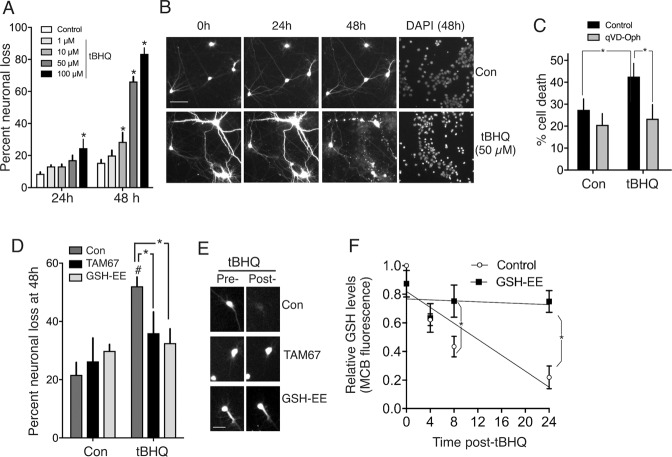
Fig. 2. TBHQ promotes Jun-dependent neurotoxicity.
A, B GFP-transfected cortical neuronal cultures17 were imaged at the indicated times post-tBHQ treatment, fixed/DAPI-stained, and viability assessed (death = cell disappearance or neurite fragmentation). *P = 0.004, 0.021, 0.0001, 0.0001; 2-way ANOVA + Dunnett’s post-hoc test, 72-121 cells analysed per condition; n = 4 independent biological replicates. C Cells were treated ± 50 µM tBHQ ± qVD-Oph (50 µM) and cell death analysed 24 h later. *P = 0.013, 0.0056, 2-way ANOVA + Sidak’s post-hoc test (n = 4 independent experiments, 481–582 cells analysed per condition). D, E Astrocyte-free neuronal cultures were transfected with eGFP plus either a control vector (ß-globin) or TAM67. The cells were treated with GSH-EE (1 mM) for 1 h where indicated, and then ±50 µM tBHQ. Cell viability was assessed 48 h later. #P = 0.0012, 2-way ANOVA + Sidak’s post-hoc test (comparing Control to tBHQ conditions). NB. No significant effect of tBHQ on cell death was observed in TAM67-transfected neurons or GSH-EE treated neurons (relative to corresponding control condition). *P (left-to-right) =0.045 and 0.013, 2-way ANOVA + Sidak’s post-hoc test, n = 6 independent experiments (130-166 cells were analysed per treatment). F Neurons were treated with tBHQ (50 µM) for the indicated times and reduced glutathione levels measured using the monochlorobimane method (See87 and “Methods”). *P = 0.0436, 0.0003, 2-way ANOVA + Sidak’s post-hoc test (n = 4).

