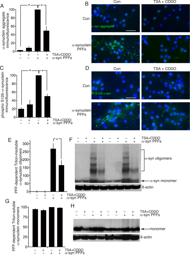
Fig. 4. CDDOTFEA and HDAC inhibition promote clearance of α-synuclein aggregates.
A–D Neuronal cultures were exposed where indicated to α-synuclein pre-formed fibrils (PFFs) on DIV4 until DIV15 to cause α-synuclein aggregate formation. On DIV15 the neurons were treated where indicated with TSA (1 µM, 8 h) followed by CDDOTFEA (250 nM, 24 h). Neurons were fixed and processed for immunofluorescence using an α-synuclein aggregate conformation-specific antibody (A, B) or a phospho-(Ser-129)- α-synuclein specific antibody (C, D) A: *P = 1.5E–09, 9.5.E–06, 1-way ANOVA + Sidak’s post-hoc test (n = 5); C: *P = 1.1E–06, 1.2E–04, 1-way ANOVA + Sidak’s post-hoc test (n = 4). B and D show example pictures relating to (A) and (C) respectively. Scale bar: 50 µm. E, F Neuronal cultures were treated as per (A–D), Triton-insoluble proteins were extracted (see “Methods”) and western analysis of oligomeric α-synuclein performed, normalized to ß-actin. *P = 0.0035, 1-way ANOVA + Sidak’s post-hoc test (n = 8). G, H Neuronal cultures were treated as per (E), except that Triton-soluble proteins were extracted (see “Methods”) and α-synuclein monomer expression quantified normalized to ß-actin (n = 8).

