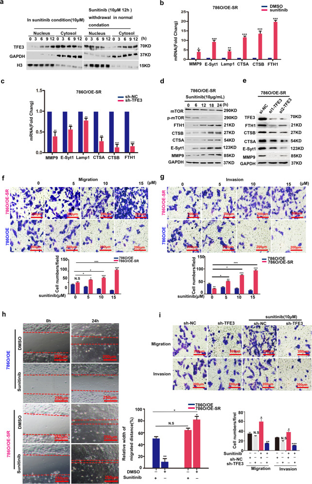
Fig. 3. Sunitinib stimulated the metastasis of 786O/OE-SR cells by activating TFE3 translocation.
a Western blot showed that expression and distribution of TFE3 was dynamic between the nucleus and the cytoplasm. GAPDH and Histone served as specific internal reference markers for cytoplasm and nuclei respectively. b The target genes of TFE3 were verified in786O/OE-SR cells treated with DMSO or sunitinib (10 μM) for 12 h. Data are shown as means ± SD. *P < 0.05, **P < 0.01, ***P < 0.001, NS: not significant. Data were obtained from three independent experiments. c After knocking down of TFE3, qPCR verified the target gene of TFE3 in 786 O/OE-SR cells treated with DMSO, sunitinib (10 μM) for 12 h. **P < 0.01, ***P < 0.001. Data are shown as means ± SD (n = 3). d Proteins downstream of TFE3 in 786 O/OE-SR cell line were upregulated with the increase in sunitinib concentration. e After knocking down of TFE3, 786 O/OE-SR were treated with sunitinib (10 μM) for 12 h cells and proteins downstream of TFE3 were down regulated. f Transwell assay to assess migration after 12 h incubation of 786 O/OE-SR with different concentration of sunitinib compared with 786 O/OE cells. Scale bar, 200 μm, ***P < 0.001, **P < 0.01, *P < 0.05, NS: not significant. Data are shown as means ± SD (n = 3). g Transwell assay to assess invasion after 12 h incubation of 786 O/OE-SR with different concentration of sunitinib compared with 786 O/OE cells. Scale bar, 200 μm. **P < 0.01, ***P < 0.001. Data are shown as means ± SD (n = 3). h Wound healing assays for 786 O/OE and 786 O/OE-SR cells with DMSO, sunitinib (10 μM) for 24 h. Scale bar, 250 μm. *P < 0.05, **P < 0.01, NS: not significant. Data are shown as means ± SD (n = 3). i Rescue experiments showed that knockout of TFE3 could reverse the effect of sunitinib on tumor metastasis in 786 O/OE-SR cells. Scale bar, 200 μm, *P < 0.05, **P < 0.01, NS: not significant. Data are shown as means ± SD (n = 3).

