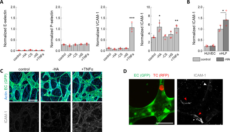
Fig. 3. Absence of endothelial activation following GCX treatment.
A Normalized (to CD31) expression of EC adhesion molecules associated with endothelial activation (n = 3 devices). B Normalized (to β-actin) expression of ICAM-1 on ECs (HUVEC) and stromal cells (nHLF) cultured in well plates after treatment with hyaluronidase (n = 3 wells). C Imaging of ICAM-1 after MVN treatment. The scale bar is 200 µm. D ICAM-1 is not highly expressed on ECs in the vicinity of TCs arrested for 6 h prior to fixing (white arrow), as compared to ICAM-1 expressed on stromal cells (red arrows). The scale bar is 60 µm. Statistical significance was assessed by student’s t test assuming normally distributed data, p < 0.05 *, p < 0.01 **, p < 0.001 ***.

