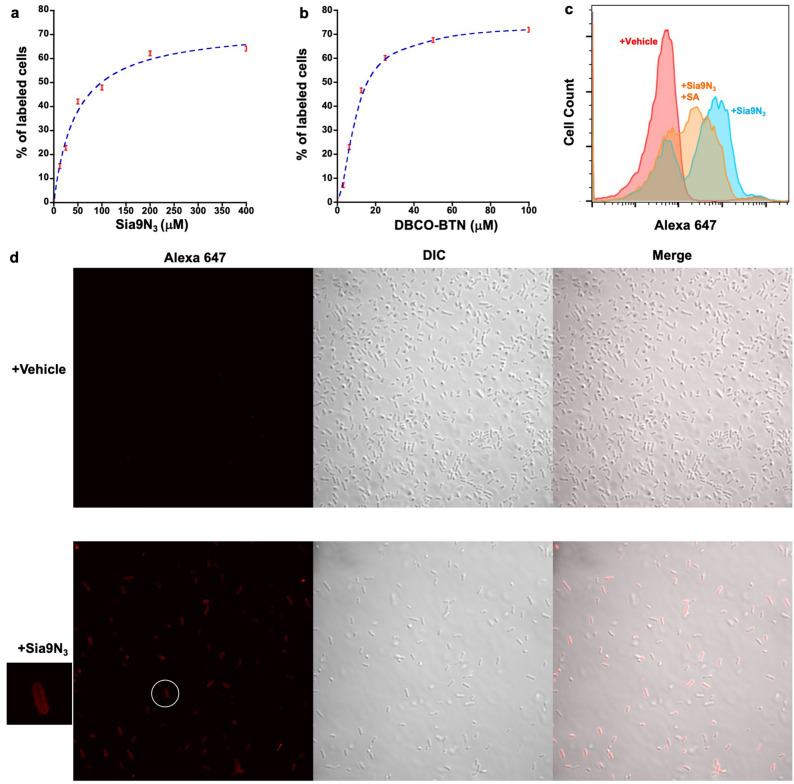
Figure 2.
Characterization of Sia9N3 induced fluorescence staining with flow cytometry and confocal fluorescence microscopy. Labeling of SA-presenting microbes is Sia9N3 (a) and DBCO-BTN (b) dose-dependent. (c) Addition of excess natural sialic acid (SA) partially competed for fluorescence labeling ([Sia9N3]: 200 μM, [SA]: 1000 μM). (d) Confocal fluorescence microscopic imaging showed that Sia9N3 induced fluorescence labeling on the bacterial cell surface.

