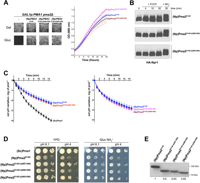
Figure 3.
The inability of C-terminally truncated PMA2 forms to activate TORC1 is not associated with reduced H+ pumping activity. (A) Left. GAL1p-PMA1 pma2Δ cells expressing, from a plasmid, either (Np)PMA2E14D, (Np)PMA2E14D-Δ(880-956), (Np)PMA2E14D-Δ(865-956), or no H+-ATPase (-) were grown for 3 days on solid medium with NH4+ as sole nitrogen source and Gal or Gluc as carbon source. Right. GAL1p-PMA1 pma2Δ cells expressing, from plasmids, either (Np)PMA2E14D, (Np)PMA2E14D-Δ(880-956), or (Np)PMA2E14D-Δ(865-956) along with HA-NPR1 were grown on Gluc NH4+ medium in a microplate reader for 28 h. Data points represent averages of the OD at 660 nm of two biological replicates; error bars represent SD. (B) The same cells as in panel A (right) were grown on Gluc NH4+ medium. After a shift to Gluc proline medium for four hours, culture samples were collected before and 4, 10, and 30 min after addition of FCCP (20 µM) or 30 min after addition of NH4+ (5 mM). Crude extracts were prepared and immunoblotted with the anti-HA antibody. The signals are from the same gel and exposure times were identical. Strains are presented in separate panels for convenience. (C) Left. GAL1p-PMA1 pma2Δ cells expressing, from plasmids, either (Sc)Pma1, (Np)PMA2E14D, or (Np)PMA2E14D-Δ(887-956) along with HA-NPR1 were grown on Gluc NH4+ medium. Acidification by these cells of the external medium was measured as described in Materials and Methods. Right. Same as in the left panel, except that cells expressing (Np)PMA2E14D, (Np)PMA2E14D-Δ(880-956), or (Np)PMA2E14D-Δ(865-956) were analyzed. Average values of three biological replicates are shown, and error bars correspond to SD. (D) GAL1p-PMA1 pma2Δ cells expressing, from plasmids, either (Sc)Pma1, (Np)PMA2E14D, (Np)PMA2E14D-Δ(887-956), (Np)PMA2E14D-Δ(880-956), or (Np)PMA2E14D-Δ(865-956) along with HA-NPR1 were spotted in two-fold serial dilutions on solid rich (YPD) or Gluc NH4+ (pH 6.1 or 4) medium and incubated for four days. (E) GAL1p-PMA1 pma2Δ cells expressing, from plasmids, either (Np)PMA2E14D, (Np)PMA2E14D-Δ(887-956), (Np)PMA2E14D-Δ(880-956) or (Np)PMA2E14D-Δ(865-956) along with HA-NPR1 were grown on Gluc NH4+ medium. After a shift to Gluc proline medium for four hours, the cells were collected. Crude extracts were prepared and immunoblotted with the anti-polyHis antibody. Direct Blue 71 staining (Sigma-Aldrich) was used for quantitative comparisons and signals were normalized to the signal of (Np)PMA2E14D-expressing cells. Original blots of figure panels B and E are presented in Fig. S3.

