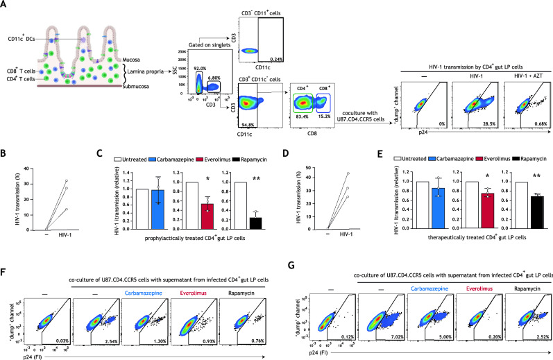
Figure 4.
Autophagy-enhancing drugs everolimus and rapamycin limit intestinal HIV-1 transmission and reduce ongoing HIV-1 replication in intestinal CD4 + T cells. (A) Human intestinal lamina propria lymphocytes (LPLs) were isolated as previously described40. Gating strategy employed to identify gut CD4 + T cells is shown; an extended gating strategy is available in Figure S7D, Supplementary Information. Following HIV-1 infection, LPLs were washed extensively to remove input virus, and subsequently co-cultured with permissive U87.CD4.CCR5 cells for 72 h. HIV-1 transmission by gut LPLs was determined by intracellular p24 staining; treatment of LPLs with HIV-1 replication inhibitor AZT (zidovudine, 20 μM) confirmed productive intestinal HIV-1 infection. (B,C) LPLs were prophylactically treated with carbamazepine (100 μM), everolimus (5 nM), or rapamycin (100 nM) for 15 h, or left untreated, followed by infection with HIV-1 NL4.3BaL for 24 h. Gut LPLs were harvested and washed to remove input virus, and subsequently co-cultured with U87.CD4.CCR5 cells. HIV-1 transmission by LPLs was assessed in LPL-U87.CD4.CCR5 co-culture for 72 h, determined by intracellular p24 staining by flow cytometer. T cell-marker CD3 was used to exclude single LPLs and LPL-U87.CD4.CCR5 conjugates from analysis. A detailed graphical depiction of HIV-1 transmission by gut LPLs is available in Supplementary Information Figure S7E. Data are mean ± SE of n = 3 donors measured in duplicate. Open circles represent the mean of duplicates from each independent experiment. Untreated, HIV-1 infected cells was set at 1, *P < 0.05, **P < 0.01, one-sample t test. (D,E) LPLs were infected with HIV-1 NL4.3BaL for 15 h, and subsequently therapeutically treated with carbamazepine (100 μM), everolimus (5 nM), or rapamycin (100 nM) for 24 h, or left untreated. Gut LPLs were harvested and washed to remove input virus, and subsequently co-cultured with U87.CD4.CCR5 cells. HIV-1 transmission by LPLs was assessed in LPL-U87.CD4.CCR5 co-culture for 72 h, determined by intracellular p24 staining by flow cytometer. T cell-marker CD3 was used to exclude single LPLs and LPL-U87.CD4.CCR5 conjugates from analysis. A detailed graphical depiction of HIV-1 transmission by gut LPLs is available in Figure S7F, Supplementary Information. (F) Following prophylactic treatment and infection with HIV-1 NL4.3BaL (as in B,C), LPLs were extensively washed to remove input virus and replated in new medium in a 96-well plate. (G) Following infection with HIV-1 NL4.3BaL and therapeutic treatment (as in D,E), LPLs were extensively washed to remove input virus and replated in new medium in a 96-well plate. (F,G) Supernatants from infected intestinal prophylactically—(F) or therapeutically—(G) treated LPLs were collected 24 h after replating and co-cultured with U87.CD4.CCR5 cells for 72 h, to further confirm productive intestinal HIV-1 infection. HIV-1 infection of U87.CD4.CCR5 cells was determined by intracellular p24 staining by flow cytometer. Representative (n = 2 tissue donors) flow cytometry plots of HIV-1 infection of U87.CD4.CCR5 cells incubated with supernatants from HIV-1 infected gut LPLs.

