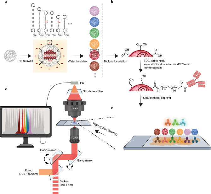
Fig. 1. Schematics of preparing Rdots and their biofunctionalization.
a 20 nm polystyrene (PS) nanoparticles are first swelled in organic solvents such as tetrahydrofuran (THF), and small-molecule Raman probes are allowed to diffuse into the nanoparticles, followed by shrinking the nanoparticles to entrap the Raman probes to generate Rdots. The dense packing of Raman probes inside nanoparticles makes Rdots ultra bright. b For easy biofunctionalization, Rdots are first conjugated with a mixture of amine-PEG8-alcohol and amine-PEG16-acid through EDC/NHS coupling to react with abundant carboxyl groups on the surface. Such combination of the two PEG chains can help reduce the non-specific binding and aggregation. Then IgG or other amine bearing bio-molecules are conjugated to the carboxyl groups from PEG-acids. This two-step procedure helps to increase the hydrophilicity and to greatly reduce non-specific binding. c Simultaneous immunostaining of Rdots and fluorescence probes (shown in shaded antibodies). d Multiplexed imaging with stimulated Raman scattering (SRS) microscopy, thanks to the narrow peak width of Rdots.

