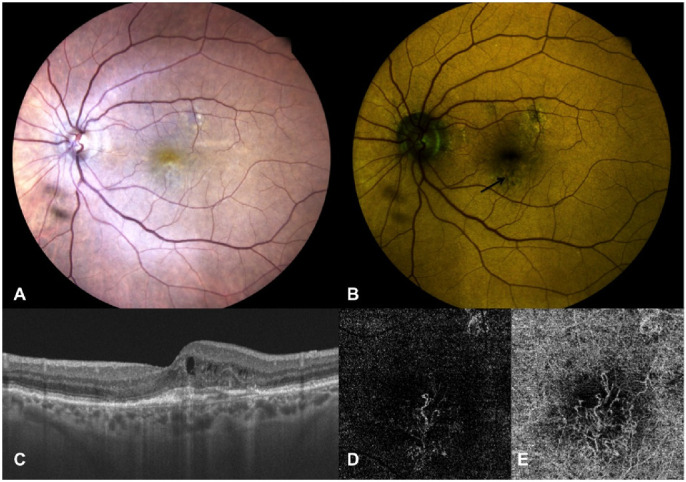
Figure 3.

Left eye of an 85-year-old female patient affected by active MNV secondary to AMD. (A) True-color fundus photography showing a whitish-yellowish central area. (B) Color FAF showing irregular areas of altered autofluorescence surrounding the fovea (indicated by black arrow), with higher intensity of the quantitatively evaluated emitting components in red (REFC intensity 71.9, GEFC intensity 57.4) at a mean wavelength of 579.09 nm. (C) OCT vertical B-scan centered on the fovea showing thickened subfoveal outer retina layers with overlying small intraretinal cysts and initial outer retina tubulations; no SRF is detectable. (D, E) OCT-A of the outer retina (D) and choriocapillaris (E) slabs showing a neovascular net interesting both slabs but with larger size of the MNV detected at the choriocapillaris.
