Figure 4.

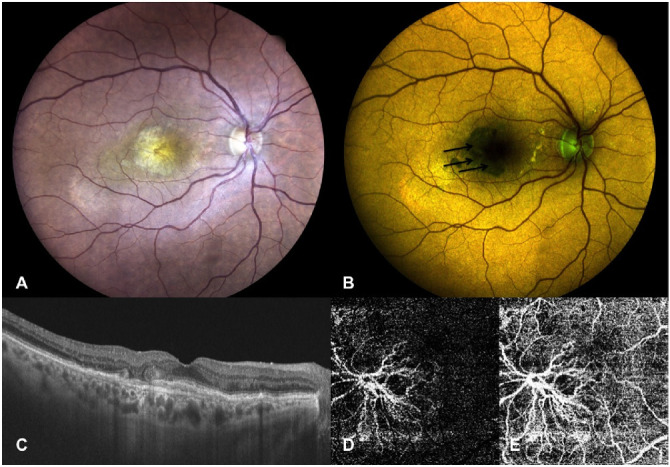
Right eye of a 77-year-old female patient affected by inactive MNV secondary to AMD. (A) True-color fundus photography showing presence of a fibrovascular lesion. (B) Color FAF showing reduced intensity of the quantitatively evaluated emitting autofluorescence components (REFC intensity 24.1, GEFC intensity 22.5) at a mean wavelength of 588.17 nm (indicated by black arrows). (C) OCT horizontal B-scan centered on the fovea showing thickened subfoveal outer retina layers with no signs of MNV activity (SRF or IRF) and perifoveal outer retina tubulations. (D, E) OCT-A of the outer retina (D) and choriocapillaris (E) slabs showing a large neovascular net interesting both slabs.
