Figure 1.
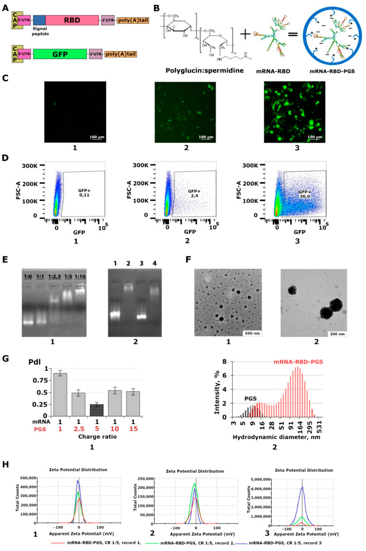
Preparation and characterization of mRNA and polyglucin:spermidine conjugate (PGS) complexes. (A) Schematic representation of mRNA-RBD, and mRNA-GFP. mRNA has a cap, poly (A) tail, 3’ and 5’ UTRs, and ORF encoding receptor binding domain (RBD) or green fluorescent protein (GFP). mRNA-RBD additionally contains a signal peptide to increase the secretion of the synthesized protein. (B) Self-assembly of mRNA-RBD-PGS or mRNA-GFP-PGS complexes. The chemical formula of the polyglucin:spermidine conjugate and the hypothetical structure of mRNA and mRNA-PGS complexes is presented. (C) Transfection of HEK293T cell culture with: (1) mRNA-GFP, (2) mRNA-GFP-PGS, and (3) mRNA-GFP-Lipofectamine 3000. Results were visualized with an Olympus CKX53 microscope 24 h after transfection. (D) Flow cytometry analysis of GFP expression in transfected cells: (1) mRNA-GFP, (2) mRNA-GFP-PGS, and (3) mRNA-GFP-Lipofectamine 3000. Results were visualized 24 h after transfection. (E) Characterization of mRNA-RBD-PGS complexes: (1) Electrophoresis in 2% agarose gel. Selection of the mRNA:PGS ratio. The charge ratios are indicated above the track, (2) Electrophoresis in 2% agarose gel. Control of the degree of coverage of mRNA with PGS conjugate at N:P equal to 1:5 before immunization. mRNA encapsulated with PGS loses its mobility in an electric field. 1—mRNA-RBD, 2—mRNA-RBD-PGS, 3—mRNA-GFP, 4—mRNA-GFP-PGS. (F) Electron micrograph of mRNA-RBD-PGS complexes at N:P equal to 1:5, (1, 2). (G) Dynamic light scattering: (1) polydispersity index (PdI), (2) size distribution profiles of nanoconstructions formed by mRNA-RBD upon complexation with PGS. (H) Measurement of the zeta potential of the resulting complexes at N:P equal to 1:5 in three different series (1,2,3).

