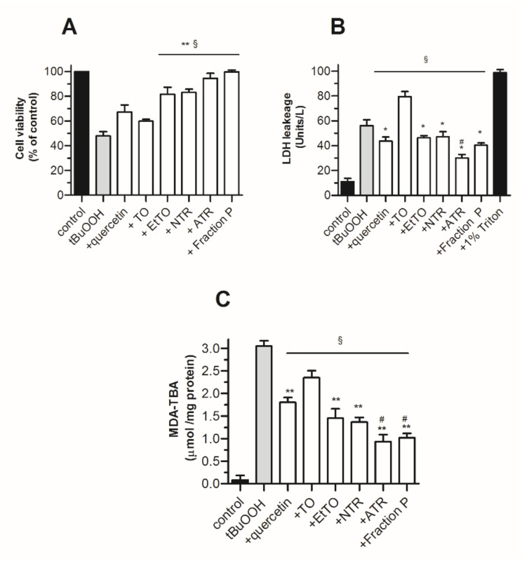
Figure 3.
Protective effect of TO extracts and metabolites against tBuOOH-induced cytotoxicity in 3T3 fibroblasts (1 × 10⁶ cells/mL). Cell viability and oxidative stress were assessed after 4 h of incubation in the absence or presence of test compounds at 15 µg/mL, by (A) LDH leakage in culture supernatant, (B) neutral red-based viability assay, (C) MDA-TBA levels. Quercetin was used at 3 µg/mL. Values represent means ± SEM (n = 8). Statistics: * p < 0.05 and ** p < 0.01 vs. TO, # p < 0.05 vs. EtTO and § p < 0.01 vs. tBuOOH, by one-way ANOVA (p < 0.01) followed by Newman–Keuls test.

