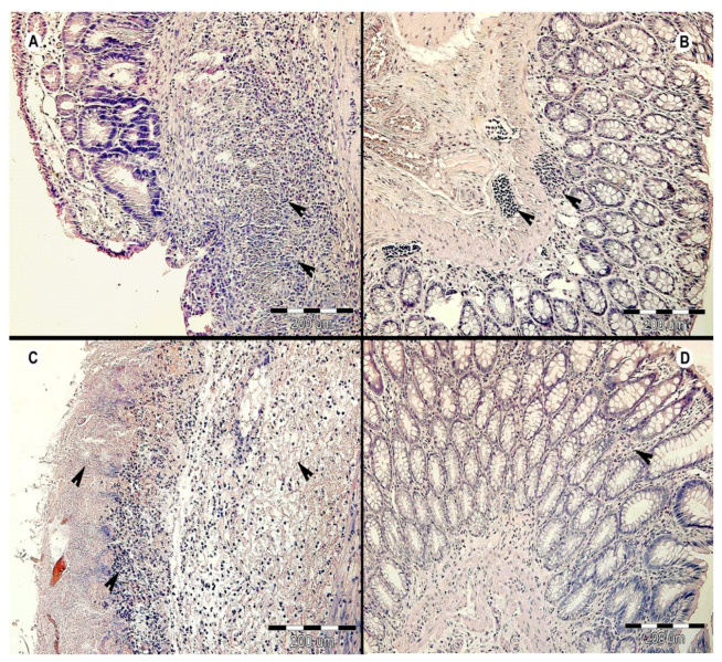
Figure 3.
Histopathological examination of the large intestine of rats on Day 31 of the experiment. The representative cross sections are displayed from each experimental group: (A) AC group (i.e., antibiotic- and colitis-treated), black arrows indicate inflammatory infiltrate in mucosa and submucosa within necrotic tissue; (B) HA group (i.e., colonized and antibiotic-treated), black arrows indicate focal inflammatory infiltrate; (C) HC group (i.e., colonized and colitis-treated), black arrows—upper left indicates necrosis of mucosal layer, lower left indicates lymphoplasmacytic inflammatory infiltrate, and right indicates submucosal edema; (D) HAC group (i.e., colonized, antibiotic-, and colitis-treated), black arrow indicates inflammatory infiltrate.

