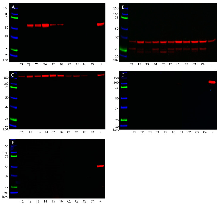
Figure 7.
Western blotting of protein extracted from six metastatic head and neck cutaneous squamous cell carcinoma (mHNcSCC) tissue samples (T1–T6) and four mHNcSCC-derived primary cell lines (C1–C4) detected the expression angiotensinogen (A) in five of the six tissue samples at the appropriate molecular weight of 55 kDa, but not in any of the mHNcSCC-derived primary cell lines. PRR (B) was detected in all of the six tissue samples and four primary cell lines at the expected molecular weight at 35 kDa for the full-length transmembrane isoform and the shorter secreted isoform. ACE (C) was detected in all 6 tissue samples, but only three of the four cell lines, at the appropriate weight of 195 kDa. ACE2 (D) and AT2R (E) were not detected in any of the six tissue samples or four primary cell line samples investigated, but were present in the positive controls. Lanes 1–6 indicate six tissue samples used, lanes 7–10 indicate cell lines. +ve indicates positive control: plasma for angiotensinogen; tonsil for PRR; mouse lung for ACE; kidney for ACE2; and mouse heart for AT2R. The molecular weight ladder (kDa) is labeled for each blot. Blots for α-tubulin are presented in Figure S7.

