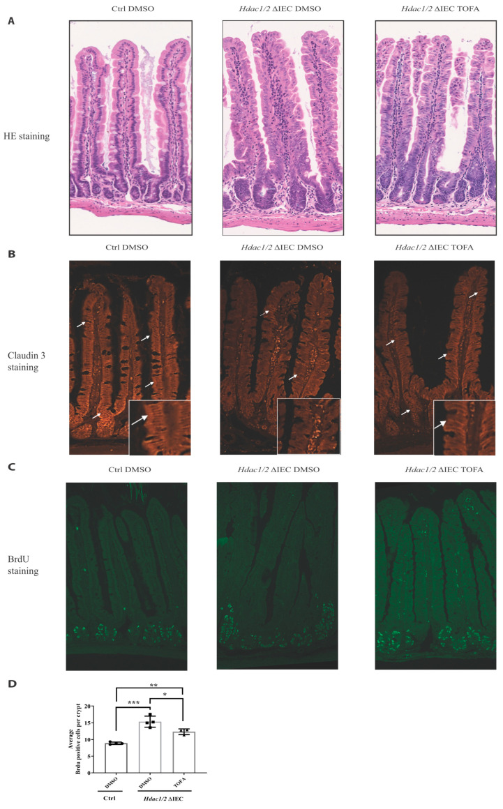
Figure 5.
Tofacitinib treatment ameliorates intestinal architecture and decreases intestinal epithelial cell proliferation in villin-Cre Hdac1 and Hdac2 deficient mice. Jejunal tissue sections, from wild-type (Ctrl) and villin-Cre Hdac1−/−; Hdac2−/− mice (Hdac1/2 ΔIEC) treated without or with Tofacitinib (DMSO or TOFA) for five days, were stained with hematoxylin and eosin (HE) (A) (Magnification: 10×) or were stained with an antibody against claudin 3 (n = 3) (white arrows) (B) (Magnification: 20×). (C) Wild-type (Ctrl) and villin-Cre Hdac1−/−; Hdac2−/− mice, treated without or with Tofacitinib (DMSO or TOFA) for five days, were injected with BrdU for 2 h. Jejunal tissue sections were stained with an antibody against incorporated BrdU. Magnification: 10×. (D) The average number of BrdU-labeled proliferative cells per jejunal crypts was measured (n = 3, 20–30 crypts each). Results represent the mean ± SEM (* p ≤ 0.05; ** p ≤ 0.01; *** p ≤ 0.005).

