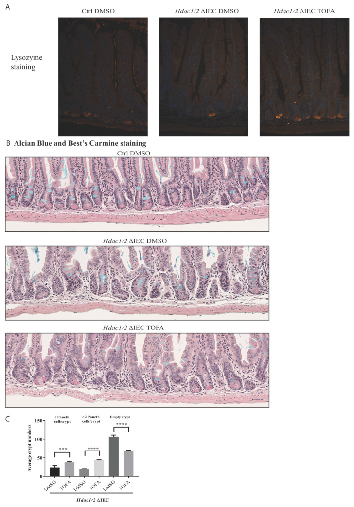
Figure 6.
Tofacitinib treatment increases the number of Paneth cells in villin-Cre Hdac1 and Hdac2 deficient mice. Jejunal tissue sections, obtained from wild-type (Ctrl) as well as villin-Cre Hdac1−/−; Hdac2−/− mice (Hdac1/2 ΔIEC) treated without or with Tofacitinib (DMSO or TOFA) for five days, were stained with an antibody against lysozyme for Paneth cell staining (white arrows), and merged with DAPI nuclear staining (A), or with Best’s Carmine for Paneth cell staining (black arrows) and Alcian Blue for goblet cell staining (B). Magnification 10×. (C) The average number of crypts with none, or with one, two or more Paneth cells was compared between jejunal tissue sections from villin-Cre Hdac1−/−; Hdac2−/− mice treated without or with Tofacitinib (DMSO or TOFA) (n = 3, 150 crypts each). Results represent the mean ± SEM (*** p ≤ 0.005; **** p ≤ 0.001).

