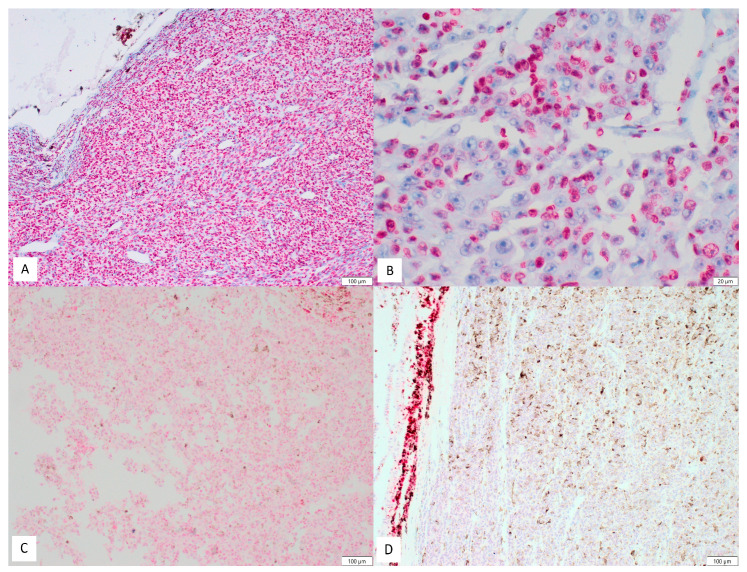
Figure 1.
Examples of PARP-1 staining in uveal melanoma cells: (A) example of high expression (100×); (B) example of high expression—intense nuclear staining visible only in about half of tumoral cells (400×); (C) example of low expression (100×); and (D) complete lack of staining in tumoral cells. Focally positive internal control (100×).

