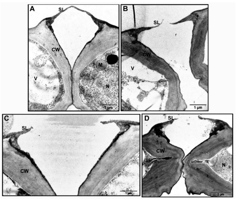
Figure 8.
TEM of stomata complex and GCs (transdermal sections; adaxial side) from rosette leaves of WT, tgg1, tgg2 single, and tgg1 tgg2 double mutants of Arabidopsis revealed variations for outer stomatal ledge and cell wall. (A). WT: Stomata complex showing nucleus, mitochondria, and vacuole in either of the GC. Cell wall is differentially thickened with thickest near the stomatal pore, and thin between the GCs. The stomatal ledge is attached at the end of stomatal pore. (B). tgg1 single mutant: Stomata complex showing vacuolated GC, with thick cell wall near the stomatal pore. GCs showing stomatal ledges at the end of stomatal pore. (C). tgg2 single mutant: Stomata complex showing thick cell wall and reduced stomatal ledges at the end of stomatal pore. (D). tgg1 tgg2 double mutant: Stomata complex showing very thick cell wall between the GCs and very thick stomatal ledges. CW = cell wall, M = mitochondria, N = nucleus, SL = stomatal ledge, and V = vacuole (Bars in A–C = 1 μm and in D =2 μm), (Magnification = 18,500× in A–C; and 11,000× in D).

