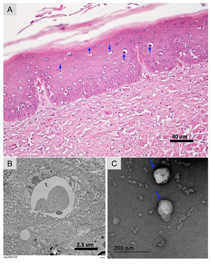
Figure 1.
Pathological and transmission electron microscopic analysis of cutaneous tissue collected from a wild green sea turtle. (A) Histological changes are characterized by vacuolation and intracytoplasmic eosinophilic inclusions in cells in the stratum spinosum with frequent displacement of the nucleus to the periphery (blue arrows). H&E stain, scale bar = 40 µm. (B) Transmission electron microscopy (TEM) on tissue section, no viral particles were discerned in these inclusions, which appeared to be composed of proteinaceous material. Scale bar = 2.5 µm. (C) Cheloniid poxvirus particles showing brick-shaped virion with regularly spaced thread-like ridges comprising the exposed surface, measuring approximately 140 nm × 98 nm. Outer envelope of cheloniid poxvirus particles is highlighted with a blue arrow.

