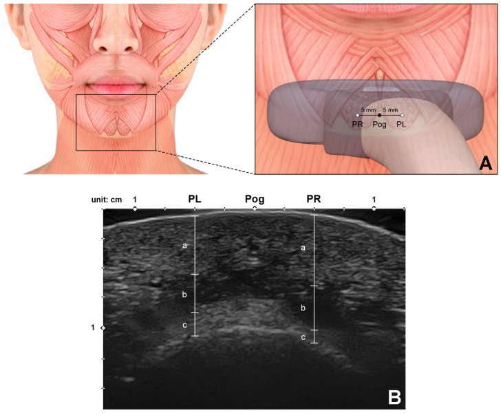
Figure 7.
Ultrasonographic analysis of the mentalis muscle. (A) Reference points for observing and measuring the thickness of the mentalis muscle. Pog, pogonion; PL, the point 5 mm left from the Pog; PR, the point 5 mm right from the Pog. A linear probe was applied horizontally at the Pog perpendicular to the skin surface to obtain the ultrasonographic image. (B) Ultrasonographic image and the parameters measured for the mentalis muscle and surrounding tissues (B mode, transverse view, 15-MHz linear transducer). Each parameter was measured at both PL and PR.; a, the depth of the mentalis muscle below the skin surface; b, the thickness of the mentalis muscle; c, the distance from the bone to the mentalis muscle were measured.

