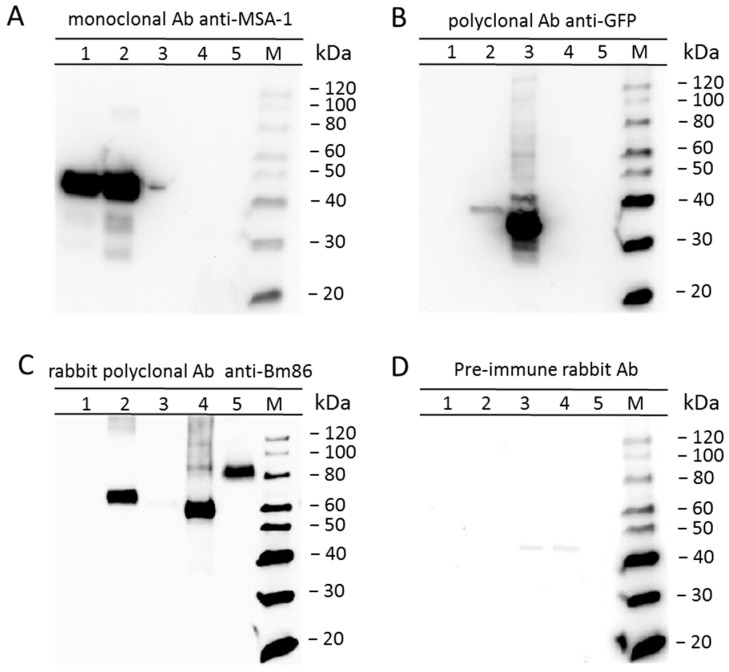
Figure 4.
Expression of Bm86Ch and eGFP by B. bovis/Bm86/eGFP demonstrated by Western blot analysis: numbers 1 to 5 indicate the lysate of B. bovis S74-T3Bo, the lysate of transfected B. bovis/Bm86/eGFP, the recombinant eGFP-BSD, the recombinant Bm86, and the R. microplus midgut, respectively. (A) Monoclonal antibody (Ab) anti-MSA-1 Babb35. (B) Rabbit polyclonal Ab anti-GFP. (C) Rabbit polyclonal Ab anti-Bm86. (D) Pre-immune rabbit serum. The protein molecular weight marker (M) is shown on the right side of each immunoblot.

