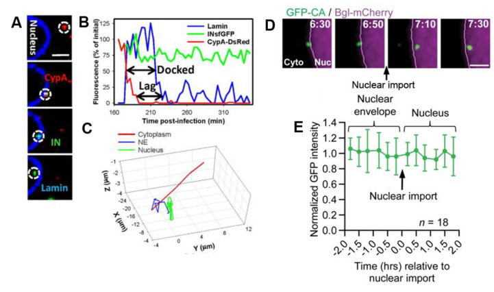
Figure 4.
Two conflicting examples of uncoating studies based on live-cell imaging of the loss of capsid labeling. (A) Imaging of uncoating at the nuclear pore by time lapse imaging of HeLa-derived cells expressing EBFP2-Lamin and infected with INsfGFP/CypA-DsRed-labeled HIV-1 pseudoviruses. The red signal (CypA-DsRed) is lost upon nuclear entry. (B) Fluorescence intensity time trace and (C) trajectory of a viral particle revealing the capsid loss during docking at the nuclear envelope characterized by confined movements (reproduced with permission from [13], copyright 2018, Cell Press). (D) Time lapse images of CA-eGFP labeled viral particles in HeLa cells expressing BglG-mCherry (located in the cell nucleus) from 6.5 h.p.i. to 7.5 h.p.i. (scale bar 2 µm) (E) The normalized mean intensity time trace of CA-eGFP from the viral particle indicate that most CA-eGFP molecules remain associated with the viral core upon nuclear entry (reproduced with permission from [18], copyright 2020, PNAS).

